


Illuminating the Microscopic World: Exploring Fluorescence Sources and filters for Advanced Microscopy by different suppliers
About external fluorescence sources:
In the field of microscopy, the ability to illuminate and visualize specimens with exceptional clarity and precision is paramount. Fluorescence microscopy, a powerful technique widely used in biological, medical, and materials science research, relies on the use of fluorescence sources that can excite specific molecules within a sample to emit light of different colors. These sources play a critical role in revealing the intricate details of cells, tissues, and materials at the microscopic level, shedding light on important scientific questions. In this article, we take a journey through the diverse and evolving landscape of fluorescence sources for microscopy, exploring the technologies that enable researchers to unlock the hidden secrets of the microcosm. From traditional light sources to cutting-edge laser systems, we will illuminate the fascinating world of fluorescence microscopy and the vital role its sources play in shaping our understanding of the minute and the magnificent.
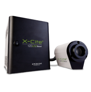
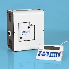
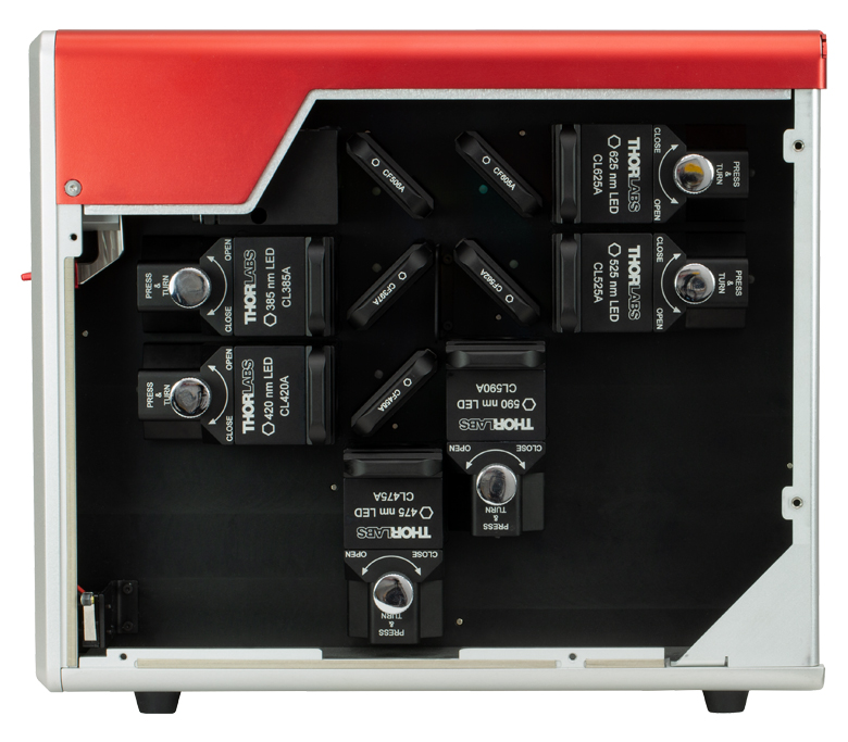
Fig. 1 – Fluorescence sources from different suppliers [1-3]
How to choose correct fluorescence light source
Choosing the right fluorescence light source for microscopy is critical to achieving optimal imaging results. Here are five key points to consider when making this important decision:
1. Wavelength range and compatibility:
Fluorescence adapters extend the capabilities of your stereomicroscope by allowing you to perform fluorescence imaging in addition to traditional brightfield observation. This versatility allows researchers to explore a wider range of applications and sample types.
2. Stability and Intensity:
Instead of investing in a dedicated fluorescence microscope, which can be costly, fluorescence adapters provide a more budget-friendly option for adding fluorescence capabilities to your existing stereomicroscope. This can result in significant cost savings for laboratories and research facilities.
3. Lifetime and maintenance:
Fluorescence adapters are designed to accommodate a variety of sample types, including live cells, tissues, and specimens. This flexibility makes them suitable for a wide range of biological and material science applications.
4. Noise and photobleaching:
Many fluorescence adapters are modular, allowing users to customize their setup by selecting specific excitation and emission wavelengths to meet their research needs. This modularity allows researchers to adapt their microscope for different experiments.
5. Application-specific considerations:
Fluorescence adapters are typically designed for easy integration with stereomicroscopes, minimizing the need for complex installation or modifications to your existing equipment. This ensures a seamless transition to fluorescence imaging.
About fluorescence filters:
In the world of microscopy, the quest for clarity and precision in imaging is never-ending. Fluorescence microscopy, a cornerstone of modern scientific research, relies on the delicate interplay of light and filters to unlock the mysteries of the microscopic universe. At the heart of this technique are fluorescence filters, unassuming yet indispensable components that selectively transmit certain wavelengths of light while blocking others. These filters are the gatekeepers of fluorescence, allowing scientists to visualize and distinguish specific molecules, organelles, and structures within cells, tissues, and materials with unprecedented detail.
Suspendisse interdum consectetur libero id faucibus nisl. Faucibus in ornare quam viverra orci sagittis eu volutpat. Vel facilisis volutpat est velit egestas. Pretium viverra suspendisse potenti nullam ac faucibus vitae. Aliquet nec ullamcorper sit amet massa ultricies mi quis hendrerit. Dolor magna eget est lorem. Erat pellentesque adipiscing commodo elit at. Neque convallis a cras semper auctor neque vitae tempus. Magna ac placerat vestibulum lectus mauris ultrices eros. Diam maecenas ultricies mi eget mauris. Feugiat sed lectus vestibulum mattis ullamcorper velit sed ullamcorper.
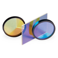
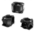
Fig. 2 – Example of fluorescence filters [4]
How to choose correct fluorescence filtres
Selecting the right fluorescence filters for microscopy is crucial to achieve accurate and high-quality imaging results. Here are five key points to consider when choosing fluorescence filters:
1. Fluorophore Compatibility:
Identify the specific fluorophores or fluorescent dyes you plan to use in your experiments. Ensure that the fluorescence filters are designed to match the excitation and emission wavelengths of your fluorophores. Opt for filters with the appropriate bandpass or longpass characteristics to effectively transmit the desired wavelengths while blocking unwanted light.
2. Filter Set Configuration:
Determine whether you need single-band or multi-band filter sets. Single-band filters are designed for imaging with a single fluorophore, while multi-band filter sets allow for the simultaneous imaging of multiple fluorophores with overlapping spectra. Choose the configuration that aligns with your imaging goals and multicolor experiments.
3. Optical Quality and Performance:
Assess the optical quality of the fluorescence filters. Look for filters that provide high transmission efficiency for excitation and emission wavelengths while minimizing autofluorescence and light leakage. High-quality filters contribute to improved signal-to-noise ratios and sharper images.
4. Sturdiness and Durability:
Consider the durability and longevity of the filters. Filters should be resistant to photobleaching and chemical damage, especially when conducting long-term or live-cell imaging experiments. Opt for filters with anti-reflective coatings that enhance their robustness and reduce stray light.
5. Microscope Compatibility:
Ensure that the selected fluorescence filters are compatible with your specific microscope model and imaging system. Verify that they fit seamlessly into the filter cubes or turret of your microscope. Consult the microscope manufacturer's recommendations or seek advice from microscopy experts to ensure compatibility.
Fig. 3 – Fluorescence supported companies by Vivanto [4]
Additionally
Additionally, consider factors such as budget constraints and the availability of filter sets from reputable manufacturers. Investing in high-quality fluorescence filters that match your experimental needs will contribute to more accurate and reliable microscopy results.
References:
[1] “X-Cite Fluorescence LED Illuminators | Excelitas.” Homepage | Excelitas, https://www.excelitas.com/product-category/x-cite-fluorescence-led-illuminators. Accessed 27 Sept. 2023.
[2] “PE-4000 | LED Light Sources | CoolLED LED Illumination System.” CoolLED, https://www.coolled.com/products/pe-4000/. Accessed 27 Sept. 2023.
[3] “6-Wavelength High-Power LED Sources.” Thorlabs, Inc. – Your Source for Fiber Optics, Laser Diodes, Optical Instrumentation and Polarization Measurement & Control, https://www.thorlabs.com/newgrouppage9.cfm?objectgroup_id=13597. Accessed 27 Sept. 2023.
[4] “Cubes, Sliders and Rings | Chroma Technology Corp.” Chroma Technology | Optical Filters, Custom & OEM Filter Design, Imaging Systems & Products, https://www.chroma.com/products/cubes-sliders-and-rings. Accessed 27 Sept. 2023.