


New Imaging Solution
INSCOPER transcends the realm of conventional microscope acquisition software. It offers a superior solution for capturing live cells, delivering not only efficiency but also ease of use. By adopting INSCOPER, research teams and core facilities optimize their microscopes, yielding a higher volume of specimen images while minimizing the time spent on microscope adjustments. Discover how INSCOPER achieves this transformation."
READ MORE ABOUT INSCOPER PRODUCTS
Inscoper Imaging Solution presents a groundbreaking hardware solution that redefines the control of fluorescence microscopes in the realm of live cell imaging. In today’s microscopy landscape, microscopes and their associated peripherals are typically linked to computers and managed through acquisition software like Metamorph, Micro-manager, LAS-X (Leica), NIS (Nikon), Olympus Stream, and Zen (Zeiss). These software applications are traditionally installed on a PC, operating within the Windows environment.
YouTube enthusiasts, don't miss our channel
Join us as we dive into the latest techniques, share insightful tutorials, and showcase captivating microscopy visuals. Subscribe today to stay updated on cutting-edge advancements and unlock the full potential of microscopy with INSCOPER.
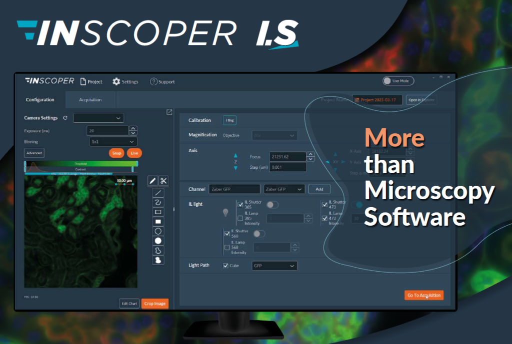
MORE THEN MICROSCOPY SOFTWARE
Inscoper I.S. is a complete, universal solution for image acquisition in fluorescence microscopy. Incorporating a specialized device controller, Inscoper I.S. provides a new user experience with improved technical performance, system integration and ease-of-use.
A Fresh Perspective
on Acquiring Images in Fluorescence Microscopy
Seamless Integration:
The Inscoper user interface is intuitively crafted to seamlessly combine various microscopy manipulations (SPIM, FRAP, high-content screening, etc.) with each other and across all time and spatial dimensions (XYZT, Θ). This imaging software effortlessly adapts to the constraints imposed by biology and photonics, offering researchers unparalleled versatility.
Universal Compatibility with Third-Party Devices:
Inscoper Imaging Software boasts full compatibility with motorized microscopes from the industry's leading manufacturers, including Leica, Nikon, Olympus, and Zeiss. It seamlessly integrates with all third-party devices and add-ons used with these microscopes, such as cameras, light sources, optical modules, microfluidic devices, and more. Even home-made equipment can be incorporated, provided its communication protocol meets a minimum documentation standard.
Precise and Reproducible Acquisitions:
Inscoper's advanced control technique synchronizes the camera and all third-party microscope devices. This synchronization guarantees the stability of acquisitions over time, eliminating jitter, and ensures complete reproducibility between sessions.
User-Centric Interface Design:
The Inscoper user interface is meticulously crafted to resemble a modern mobile app or website, offering an appealing visual experience and a user-friendly journey. Free from cumbersome drop-down menus or cluttered windows, this interface allows users to direct their focus and energy entirely toward the complexities and uncertainties inherent in studying their biological specimens.
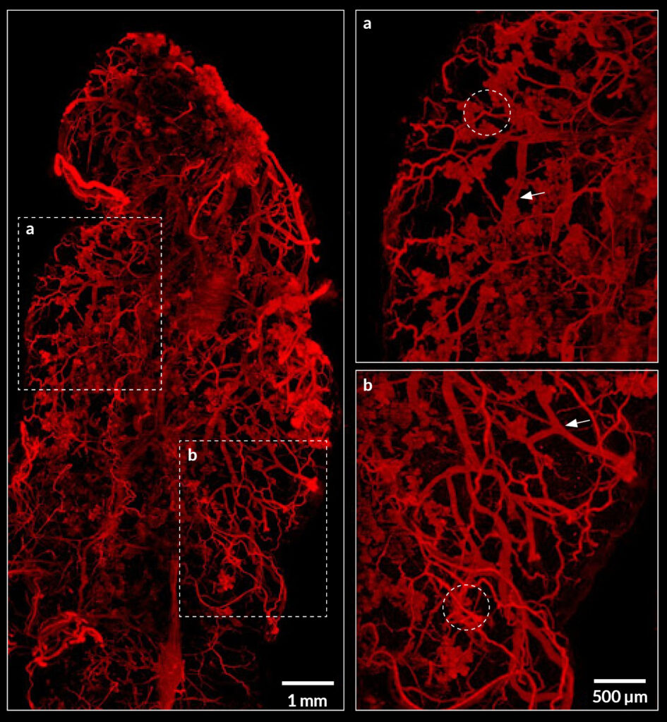
IMAGING CLEARED ORGAN WITH AN ULTRAMACROSCOPE USING INSCOPER IMAGING SOLUTION
Imaging of myoepithelial cells in cleared mammary gland using the ultramacroscope Maximum projection image of an entire iDISCO-cleared mammary gland with myoepithelial cells labeling (SMA, red). Two dashed rectangles are zoomed in at the right part of the image. Dashed circles represent some clusters of alveoli and arrows point to ducts. Scale bar: 1mm (left) and 500µm (right).
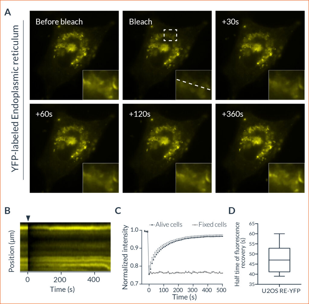
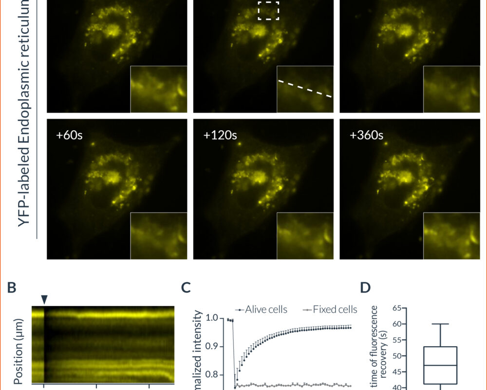
CHARACTERIZATION OF ORGANELLE DYNAMICS IN LIVING CELLS BY COMBINING OXXIUS LASERS AND INSCOPER SCANFRAP
FRAP experiment to monitor the endoplasmic reticulum dynamics in living cells (A) Representative example of HeLa cells expressing YFP-ER before and after photobleaching. The recovery of the fluorescent signal after photobleaching within the region marked by the dashed rectangle is visualized over time. This area is zoomed in at the bottom right corner of each image. The represented dashed line indicates the area used for the following kymograph. (B) Kymograph representing the evolution of the fluorescence intensity as a function of time. The black arrowhead represents the photobleaching event. (C) Normalized quantification of the fluorescence recovery following a photobleaching event (n = 7). Data are expressed according to the mean ± SEM. (D) Half-time of the fluorescence recovery in FRAP assays (n = 7).
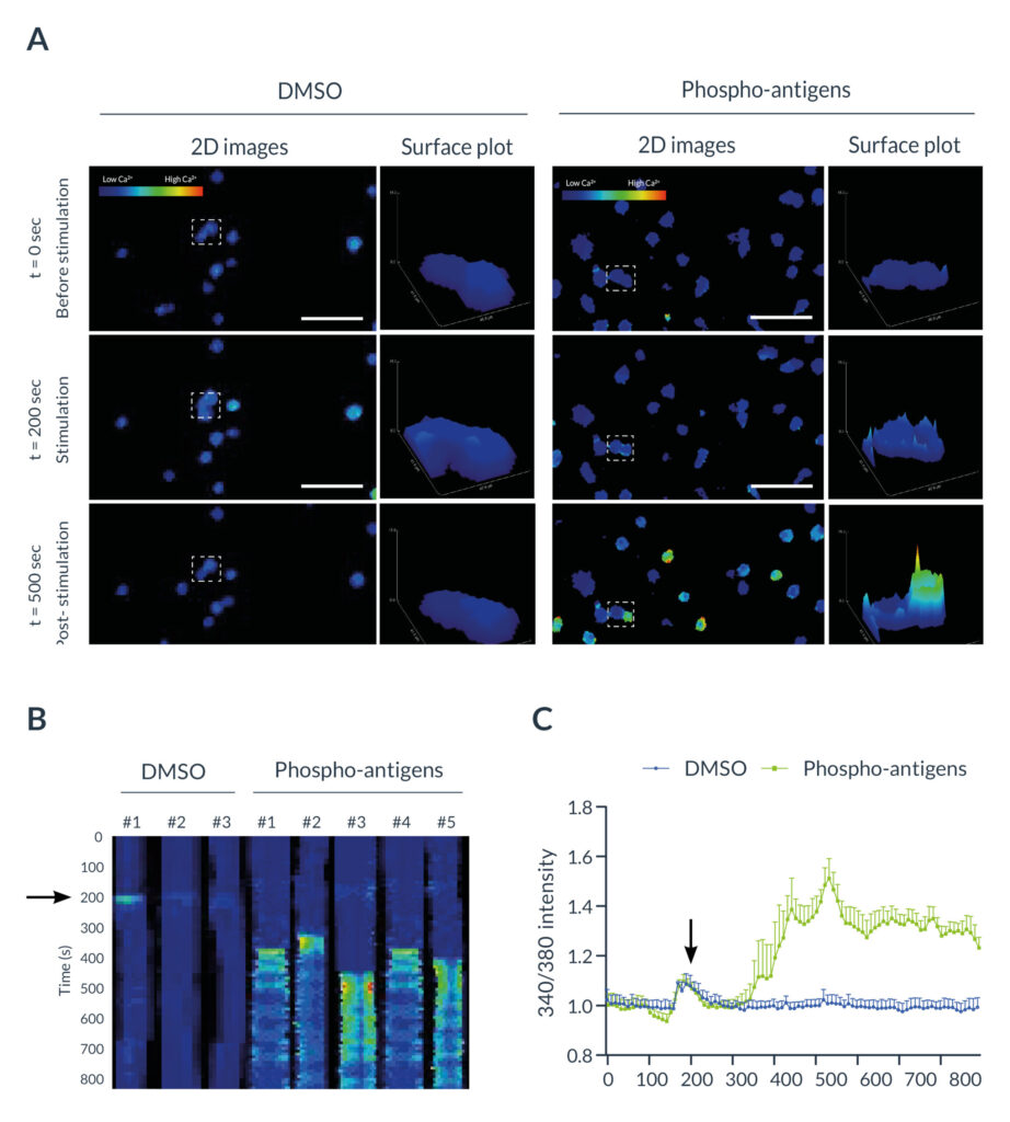
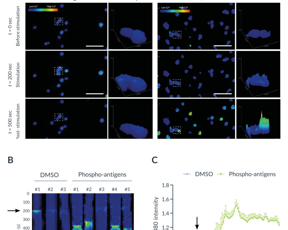
LIVE CALCIUM IMAGING TO MONITOR T CELLS ACTIVATION USING INSCOPER LIVERATIO
T cells activation monitored by ratiometric imaging with the Inscoper liveRATIO (A) Representative images of γδ T cells labeled with Fura-2 with DMSO or phospho-antigen (PA) stimulation. Surfaces plots are extracted from each dashed square. Scale bar = 100µm. (B) Kymograph representing the evolution of the fluorescence intensity as a function of time. The black arrowhead represents the addition of DMSO or PA in the medium. (C) Calcium fluctuation induced by DMSO or phospho-antigen on γδ T lymphocytes labeled with Fura-2. The black arrowhead represents the addition of DMSO or PA in the medium. Data are expressed according to the mean ± SEM.
Discover our real-world cases
We are delighted to present our collection of application and technical notes, showcasing our expertise and innovative ideas in the realm of control and image acquisition for fluorescence microscopes. Your feedback is greatly appreciated, and we encourage you to leave comments and share any items that resonate with you.
What Customers Says
Condensed Matter Physics Lab. (LPMC), Ecole Polytechnique, FRANCE
I found that the Inscoper team is a real expert with knowhows in direct control of hardware and providing customized software interface. We saved a lot of time and started the core experiments that we designed.
If your project is not the instrumentation, Inscoper could be the quickest and high-quality solution.
National Institute for Standards and Technology (NIST), Cell Systems Science Group, Gaithersburg, Maryland, USA
The Inscoper technology has become an essential part of our image collection workflow. The Inscoper controlled automated microscope operates more than 5x faster. This allows us to generate training data for deep learning-based analysis much more efficiently and allows us to perform dynamic single cell measurements with much deeper sampling than otherwise possible.
IR2 – INSERM, Institut Cochin, FRANCE
With the Inscoper solution, benefits come from the speed of acquisition. In a more global view, if we have to change the computer, the reinstallation will be easy compare to other solutions.
Simple to install, quick to learn, fast acquisition, reactive after sale service.
Inscoper clients

Are you interested?
Looking for a customized solution for your specific needs? Our team of experts are here to provide you with a solution that is a perfect fit for your needs.
Contact us today to discuss your project and discover how our customised solutions can maximise your potential for success.