


Elevating Precision and Control: Advanced Incubation Systems for Microscopy with VIVANTO support
Microscopy incubation systems are a fundamental pillar of modern scientific research, bringing new levels of precision and control to the study of living cells and organisms. These sophisticated systems provide a carefully controlled environment within the microscope that mimics the conditions necessary for cells and samples to thrive and exhibit their natural behaviour. Whether in cell biology, neuroscience or drug discovery, incubation systems have become indispensable tools, enabling researchers to delve deeper into the dynamic processes of life itself.
We explore the essential role of temperature stability, which is vital for cell and tissue viability and functionality under the microscope. We also examine how humidity control ensures that specimens remain hydrated and healthy during prolonged imaging sessions. We also discuss the importance of gas exchange capabilities when studying oxygen-sensitive processes such as cell respiration or microbial growth.
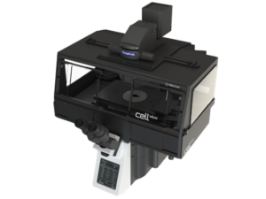
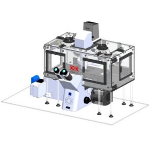

Fig. 1 – Incubation system with stage top and housing from different suppliers and for different microscopes [4].
Using advanced incubation techniques, we investigate the critical role of thermal stability, which is vital to the viability and functionality of cells and tissues under the microscope. We also observe how humidity control ensures that samples remain hydrated and healthy during long-term imaging. In many cases, we pay attention to the importance of gas exchange capabilities when studying oxygen-sensitive processes such as cellular respiration or the growth of microorganisms.
Fig. 2 – Example of OKOLAB’s stage-top incubator [3].
In summary, incubation systems in microscopy represent a vital interface between technology and biology. They enable scientists to observe the intricate dance of life at the cellular and subcellular levels, shedding light on the hidden mechanisms that govern health, disease and the natural world. As technology continues to evolve, these systems will undoubtedly continue to push the boundaries of what we can discover under the microscope, ushering in a new era of scientific enlightenment and innovation.
How to choose with key factors:
Key factors for incubation systems in microscopy include a range of considerations to ensure the controlled environment effectively supports live cell and specimen imaging. These factors are critical for maintaining the viability, health, and integrity of the samples being studied. Here are the key factors to consider when evaluating incubation systems:
1. Temperature Control:
Ensure that the incubation system provides uniform temperature distribution across the imaging area to avoid temperature gradients that could affect experimental results. Be careful with stage-top incubators.
2. Temperature Uniformity
Maintaining appropriate humidity levels is critical to prevent sample desiccation, especially during long-term imaging. Some experiments may require adjustable humidity levels to mimic specific physiological conditions.
3. Humidity Control
Maintaining appropriate humidity levels is critical to prevent sample desiccation, especially during long-term imaging. Some experiments may require adjustable humidity levels to mimic specific physiological conditions.
4. Gas Control and Exchange:
For oxygen-sensitive experiments or studies with specific gas requirements, consider incubation systems that can control and exchange gases such as oxygen, carbon dioxide and nitrogen. Gas concentration should be adjustable to suit experimental requirements.
5. Stability, precision, compatibility:
Look for systems that offer high stability and precision in temperature, humidity and gas control to ensure consistent experimental conditions. Ensure that the incubation system is compatible with your specific microscope model and can be seamlessly integrated without interfering with imaging.
6. Chamber Size and Configuration:
Consider the size and configuration of the incubation chamber. It should comfortably accommodate your sample holders, multiwell plates or other experimental setups.
7. Ease of Use:
User-friendly software and controls are essential for efficient operation. The system should allow researchers to program and monitor environmental conditions easily.
8. Fast Recovery Times:
Rapid recovery times after the microscope enclosure is opened and closed are crucial to minimize disruption to live specimens during imaging sessions.
9. Sample Accessibility:
Ensure that the incubation system permits simple access to samples for maintenance, manipulation, or addition of reagents without disturbing the environment.
10. Data Logging and Monitoring:
Choose systems that have the ability to log data, track and measure environmental conditions over time, ensuring traceability and quality control for experiments.
11. Vibration Isolation:
In certain situations, it may be necessary to isolate vibrations to prevent them from affecting the quality of images, especially in labs shared with other people.
12. Safety Features:
For safety purposes, the system must have features such as alarms or automatic shut-off mechanisms that engage when the temperature or humidity deviates from the set parameters.
Fig. 3 Example of the enclosure incubator from Tokai Hit [1]
Our supported brands
We are able to help you with these supported brands below.


Fig. 4 Our supported brands [1-4]
Final thoughts
Researchers can choose the best incubation system for their microscope experiments by evaluating factors, ensuring optimal conditions for live cell imaging and other dynamic studies.
We are available to customize a suitable solution for your needs. Contact us now to talk about your project and learn how our tailored solutions can boost your chances of success.
References:
[1] “Tokai Hit – Microscope Enclosure.” Tokaihit-Livecell, https://www.tokaihit-livecell.com/warmingbox. Accessed 22 Sept. 2023.
[2] GmbH, PeCon. “PeCon GmbH – Live Cell Imaging and Microscope Incubators.” PeCon GmbH – Live Cell Imaging and Microscope Incubators, 23 Sept. 2021, https://www.pecon.biz/.
[3] “Okolab – Home.” Okolab – Home, https://www.oko-lab.com/. Accessed 22 Sept. 2023.
[4] “Clean Enclosure for Microscopes PureBox SHIRAITO® | IVEXL.” Internet Virtual Exhibition for Life Science Industry, https://ivexl.com/en/details_service1.html?id=825. Accessed 25 Sept. 2023.
[5] Headline picture: “Ústav Molekulární Genetiky AV ČR, v. v. i.” Ústav Molekulární Genetiky AV ČR, v. v. i., https://www.img.cas.cz/. Accessed 26 Sept. 2023.
Are you interested?
Looking for a customized solution for your specific needs? Our team of experts are here to provide you with a solution that is a perfect fit for your needs.
Contact us today to discuss your project and discover how our customised solutions can maximise your potential for success.